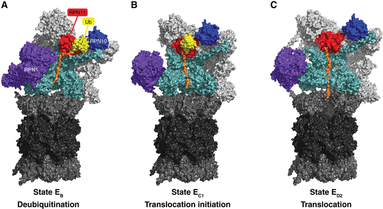
Figure 1. Conformational states of substrate-bound human proteasomes.
Cryo-EM density maps of substrate (orange) bound human proteasomes in three different stages of substrate processing are represented. A shows a conformation that is compatible with the deubiquitylation of the bound substrate. B) and C) represent two consecutive conformations of substrate translocation (EC1 and ED2) into the CP. Rpt1 and Rpt5 density maps were removed from the ATPase rings to expose the substrate translocation channel of the RP. In ED2, subunit α1 (Psma1) was also removed to show the substrate translocation channel of the CP. EB (PDB: 6MSE), EC1 (PDB: 6MSG), and ED2 (PDB: 6MSK) density maps were obtained from (Dong et al. 2018).

