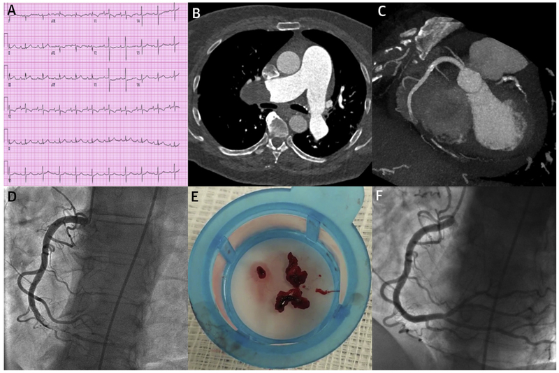
FIGURE 1. Concomitant Acute Coronary Occlusion and Pulmonary Embolism.
(A) Electrocardiogram showing a normal sinus rhythm at 62 beats/min, first degree atrioventricular block, right axis deviation, and nonspecific ST-T wave changes. (B) Computed tomography angiography showing a large clot in the right pulmonary artery. (C) Computed tomography angiography showing occlusion of the distal right coronary artery. The acute contrast cutoff along with absence of any calcification is suggestive of thrombotic occlusion. (D) Invasive coronary angiogram showing thrombotic occlusion of the distal right coronary artery. (E) Photograph of fresh thrombus retrieved with aspiration thrombectomy. (F) Invasive coronary angiogram performed after aspiration thrombectomy that shows complete resolution of the right coronary artery occlusion without any residual plaque.

