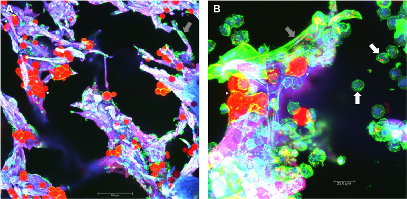
Figure 3: Example of tissue-engineered bone marrow adipose tissue.
Silk scaffolds were used as a platform to make 3D, tissue-engineered bone marrow adipose tissue (BMAT). (A) Mouse BMAT was differentiated from male, 10-month-old KaLwRij mouse-derived bone marrow mesenchymal stem cells (BM-MSCs). Briefly, BM-MSCs were seeded onto scaffolds and cultured for 6 days, then put into adipogenic media for 10 days, put into a maintenance media for 1 week, and finally imaged. Scale bar = 200 μm. (B) Mouse-derived BM-MSCs were treated as above up until adipogenesis then the mouse myeloma cell line, 5TGM1s, were co-culture with the differentiated adipocytes for 1 week in maintenance media. Scale bar = 25.0 μm. Samples were stained Oil Red O (red) and phalloidin/actin (green). The scaffolds are autofluorescent in all channels and thus appear purple. Imaging was performed with a confocal microscope using maximum projections of Z-stacked images. Many adipocytes are visible (red, white arrowheads), along with many undifferentiated stromal cells (grey arrows). Rounded green myeloma cells are seen throughout the scaffold (white arrows).

