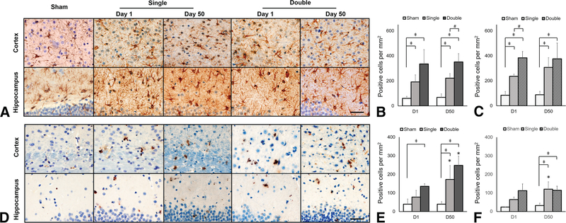
FIG 5.
Astroglial and microglial expression in the cortex and hippocampus underlying the impact region after CHI. A, GFAP-positive staining and quantification of GFAP expression in the cortex (B) and hippocampus (C) after CHI. D, CD 68 –positive staining and quantification of CD 68 expression in the cortex (E) and hippocampus (F) after CHI. As early as 24 hours after both single and double CHI, elevated GFAP is observed in both the cortex and hippocampus deep to the impact site. CD 68 expression indicating neuroinflammation is also observed at 24 hours postinjury and sustained to day 50 in the cortex after double CHI. Scale bar, 40 μm.

