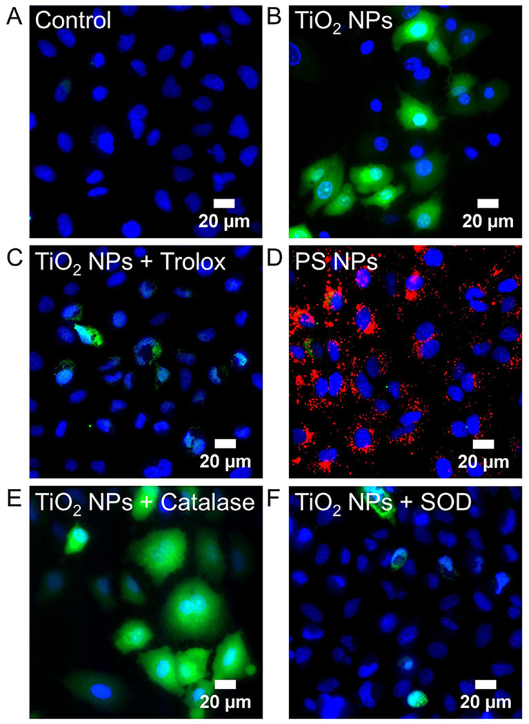
Figure 8.
H2DCF, a non-specific probe of intracellular ROS, was used to image ROS in Generation 1 and 10 A549 cells. H2DCFDA (5 μM) was incubated with cells for 30 min prior to imaging and then rinsed with PBS. A. Untreated control cells. B. TiO2 NP-treated cells (994 μg/mL, 24 hr, 37°C; Generation 1). No H2DCF signal was observed at Generation 10 (Figure S5) and all subsequent images are at Generation 1. C. Co-incubation of TiO2 NPs (994 μg/mL, 24 hr, 37°C) with Trolox (0.5 mM), a general ROS scavenger. D. Incubation with polystyrene (PS) NPs (red, 40 pM, 24 hr, 37°C). E. Co-incubation of TiO2 NPs (994 μg/mL, 24 hr, 37°C) with catalase (50 U/mL), a H2O2 scavenger. H2O2 was used as a positive control (Figure S6). F. Co-incubation of TiO2 NPs (994 μg/mL, 24 hr, 37°C) with SOD (50 U/mL), a superoxide scavenger. DAPI (blue) was used to label nuclei (50 μM, 30 min).

