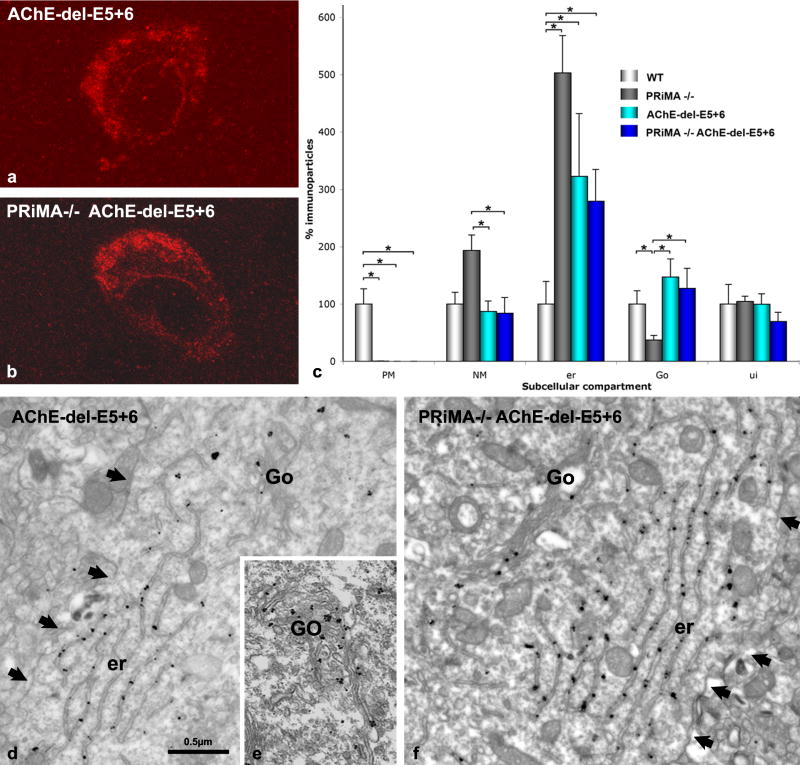
Figure 6. The AChE WAT domain targets AChE to the cell surface in WT mice and promotes retention of AChE in the endoplasmic reticulum of the PRiMA knockout strain.
(a, b) Confocal microscopy shows AChE labeling in the striatum of (a) the AChE del E5+6−/− knockout and (b) the double knockout, PRiMA−/−/AChE del E5+6−/−. Similar staining is also seen in the PRiMA−/− knockout (Fig. 3b).
(d–f) Electron microscopy shows an absence of immunoparticles at the plasma membrane of AChE del E5+6−/− (d) and PRiMA−/−/AChE del E5+6−/− (f) animals and the presence of AChE in the Golgi apparatus of the AChE del E5+6−/− mouse (e). Arrows point the plasma membrane. (c) Quantification at the EM level reveals that the reduction in the number of immunoparticles for AChE in the Golgi apparatus (Go) compared to WT animals is only seen in the PRiMA knockout, not in AChE del E5+6−/− or PRiMA−/−/AChE del E5+6−/− mice. However, immunoparticles for AChE accumulate in the endoplasmic reticulum (er) in all three knockouts. Data are analyzed as in figure 4 and are compared using the nonparametric Kruskal-Wallis one-way analysis of variance, followed by a post-hoc analysis using the Bonferroni-Dunn test.

