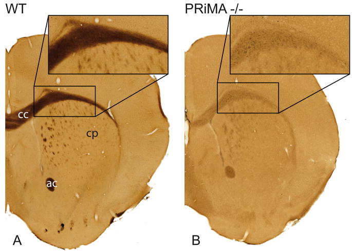
Figure 8. Histochemical detection of BChE in the anterior brain of the WT and PRiMA knockout mouse.
BChE activity in brain was revealed by thiocholine production as a brown precipitate. In the WT brain, BChE is expressed in white matter including corpus callosum (cc), white anterior commissure (ac) and white matter fascicles in the caudate putamen (cp). In the PRiMA knockout mouse, BChE labeling is markedly decreased and restricted to the cell body.

