
Figure 4.
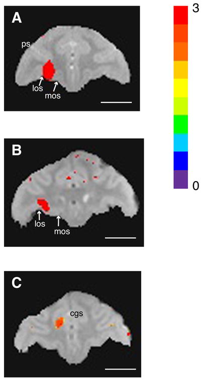
Localization of isotonic (120 mM) MnCl2 within 3 hours of injection. A. Orbitofrontal injection site in monkey T. B. Orbitofrontal injection site in monkey S. Orbitofrontal injections were localized to the lateral half of area 13 in both monkeys, with some extension into the overlying white matter. los: lateral orbital sulcus; mos: medial orbital sulcus; ps: principal sulcus. C: Anterior cingulate injection site in monkey S. The injection site was located ventral to the cingulate sulcus in area 24c. All images acquired at 4.7T with 0.5 mm3 resolution, and sequence parameters: TR100 msec, TE 3.5 (monkey S) or 4.7 msec (monkey T), apparent flip angle 45°. Mn2+ signal intensity maps are superimposed on images from separate volumes acquired with sequence parameters designed to highlight gray-white matter contrast and anatomical landmarks (TR 100 msec, TE 6.5 msec, apparent flip angle 10°). Highlighted voxels are those with signal intensities at least two standard deviations from the pre-injection mean (color bar from 0 to 3 standard deviations). cgs: cingulate sulcus. Length of all scale bars = 10 mm.