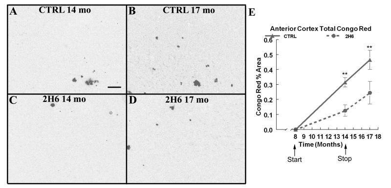
Figure 3. CONGO RED HISTOCHEMISTRY REPRESENTING COMPACT AMYLOID DEPOSITS ARE REDUCED AFTER 6 MONTHS OF SYSTEMIC ANTI-Aß ANTIBODY ADMINISTRATION AND THESE REDUCTIONS ARE MAINTAINED 3 MONTHS AFTER THE CESSATION OF TREATMENT.
Panels A & B show Congo red histochemistry in the frontal cortex of Tg2576 mice that received control antibody at 14 months of age and at 17 months of age. Panel C & D show Congo red histochemistry in Tg2576 mice that received anti-Aß antibody at 14 months of age and at 17 months of age. Scale bar = 120μm. Panel E shows quantification of the percent area occupied by Congo red positive plaques in the frontal cortex. The solid line shows the value for APP transgenic mice that received control antibody. The dotted line shows the values for APP transgenic mice that received anti-Aß 2H6 antibody. ** indicates P<0.01.

