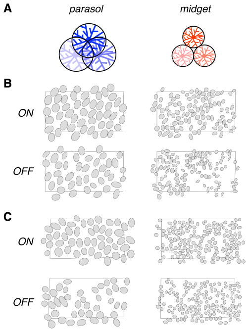
Figure 1.
Parasol and midget RF mosaics and anatomical prediction. A. Previous anatomical findings indicate that parasol cell dendritic fields overlap substantially, with the tips of each dendritic field reaching the soma of its neighbors in the mosaic, while midget cell dendritic fields abut at their boundaries. B. Each panel shows the RFs of simultaneously recorded ON and OFF parasol and midget cells from one retina, with each RF represented as the 1 standard deviation boundary of a Gaussian fit to the RF center. Black rectangles indicate the outline of recording array (1800 by 900 micrometers). Gaps in the mosaic probably represent unrecorded cells. Retinal temporal equivalent eccentricity: 6.4 mm. C. Same as in B for a second preparation (temporal equivalent eccentricity 9.0 mm).

