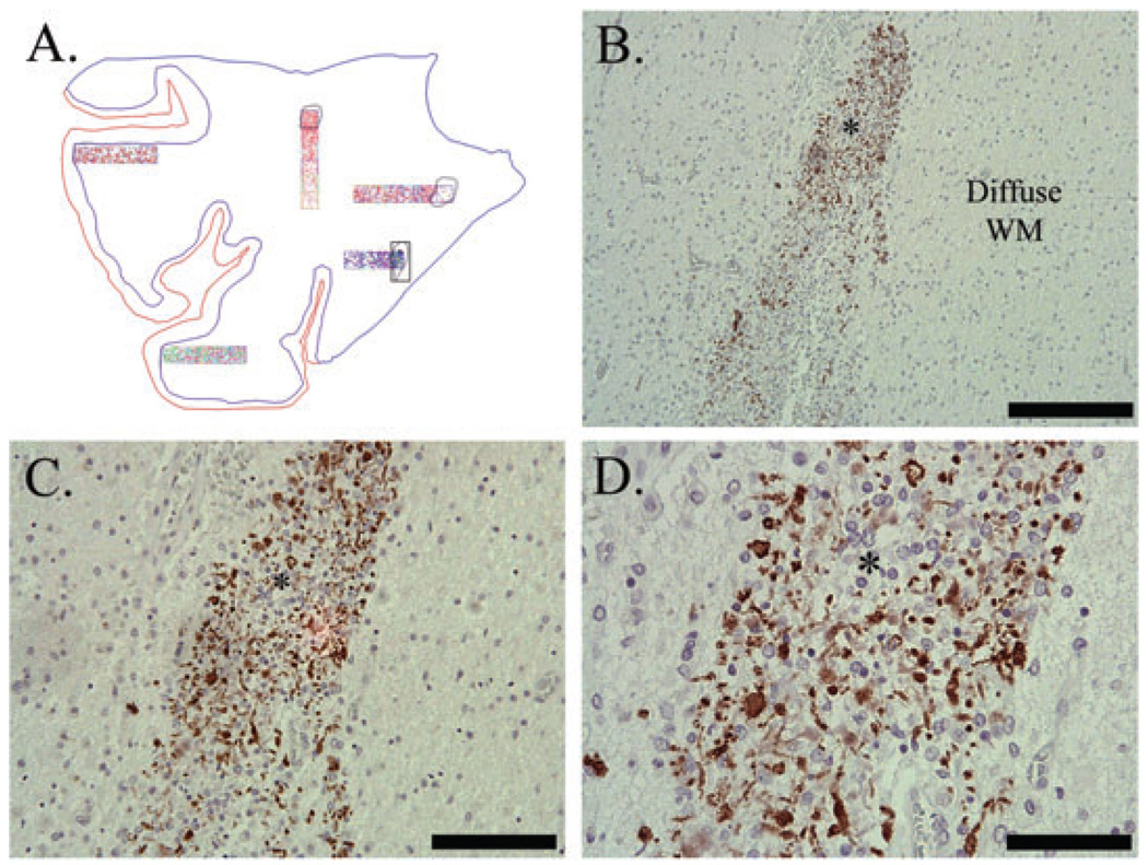
Figure 7.
Abnormal myelin basic protein (MBP) immunostaining in Case 11 (40 post-conceptional weeks) with a chronic (glial scar) necrotic focus (*). Abnormal MBP immunostaining was in the form of globular segments (lines), rare tubules (arrowhead) and rare sheaths (arrows) (D). Note that the surrounding diffuse white matter (WM) is devoid of any MBP immunostaining (B–D). Image A is the Neurolucida image of this case; necrotic focus is highlighted. Image B is taken at 100×, scale = 200 µm; C is at 200×, scale = 100 µm; D is at 400×, scale = 50 µm.

