
Figure 3.
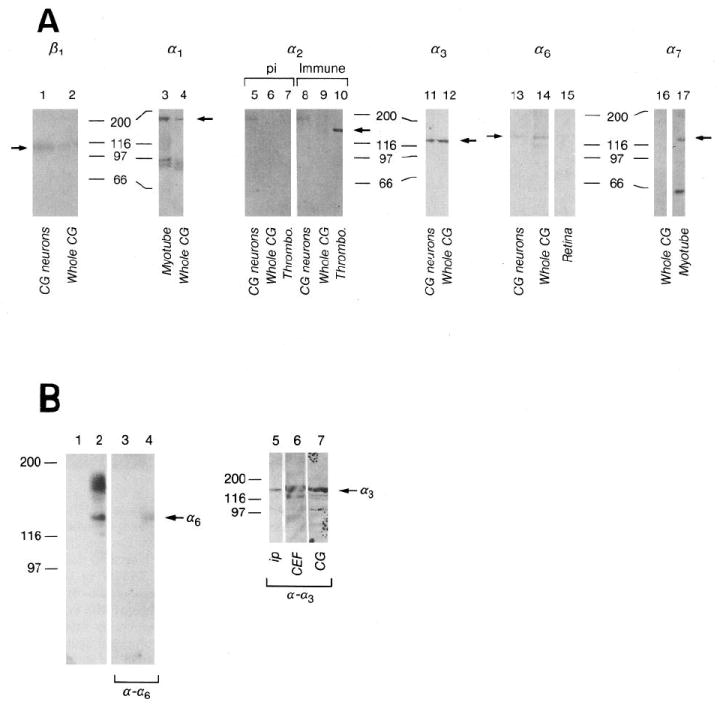
A, Integrin expression by E7-8 CG and purified CG neurons cultured on laminin-1. Whole CG (lanes 2, 4, 6, 9, 12, 14), CG membranes (lane 16), CG neurons plated on laminin-1 (lanes 1, 5, 8, 11, 13), E11 myotubes (lane 3), E11 myotube membranes (lane 17), thrombocytes (lanes 7, 10) and E6–8 retinae (lane 15) were extracted in sample buffer (lanes 1, 2, 5–17) or 1% Triton X-100 (lanes 3, 4) as described in Materials and Methods. Approximately equal amounts of protein were loaded per lane for each blot. Blots were incubated with polyclonal antibodies against α1 (lanes 3, 4), α2 cyto (preimmune, lanes 5, 6, 7; immune, lanes 8, 9, 10), α3 (Ex2, lanes 11, 12), α6 (Ex, lanes 13, 14, 15), or monoclonal antibodies against β1 (TASC, lanes 1, 2) or α7 (H1, lanes 16, 17). Immunoreactivity was visualized with alkaline phosphatase-conjugated anti-rabbit or anti-mouse antibodies. Arrows indicate bands corresponding to each subunit. The doublets around 97 kDa in lanes 3 and 4 are probably breakdown products of α1 (Duband et al., 1992). The band at 190 kDa in lanes 5 (preimmune) and 8 (immune) is nonspecific. The lower Mr band in lane 17 is a common breakdown product of α7 (Bao et al., 1993). Numbers denote positions of Mr marker proteins in kilodaltons. pi, Preimmune. B, α6 and α3 heterodimerize with β1 in CG neurons. β1-containing integrin heterodimers were immunoprecipitated from surface-biotinylated CG cells with anti-β1 (W1B10) mAb coupled to protein A–Sepharose. Immunoprecipitates were visualized directly by chemiluminescent detection of HRP–streptavidin (lane 1, protein A–Sepharose control; lane 2, antibody-coupled Sepharose), then probed with anti-α6Ex (lanes 3, 4). Similar immunoprecipitates were probed with anti-α3Ex2 (ip, lane 5). Triton X-100 extracts of chick breast fibroblasts (CEF, lane 6) and whole ciliary ganglia (CG, lane 7) were also blotted as positive controls. Arrows indicate bands corresponding to α6 or α3. Numbers indicate Mr in kilodaltons.