
Figure 4.
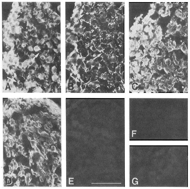
CG neurons ex press α6, α3, and β1, but not α2 or α7, in situ. Cryostat sections of E7-8 ciliary ganglia were stained with anti-integrin antibodies as described in Materials and Methods. A and B show a double-labeled section incubated with anti-β1, mAb (TASC, A) and a polyclonal antibody to neurofilament (B). Anti-β1 immunoreactivity appears to be highly coincident with that of the neuronal marker, notably in structures which appear to be axons (arrows). Other sections were incubated with antibodies to α3 (cytoplasmic domain A-form, C), α6 (D), α6 preimmune IgG (E), α2 (cytoplasmic domain, F), and α7, mAb (G). As for β1, α3 and α6 immunoreactivity often colocalizes with that of neuronal markers (data not shown). The α2 and α7 antibodies were immunoreactive with control tissues (epithelium and muscle, respectively) in the same experiments (data not shown). Scale bar, 100 μm.