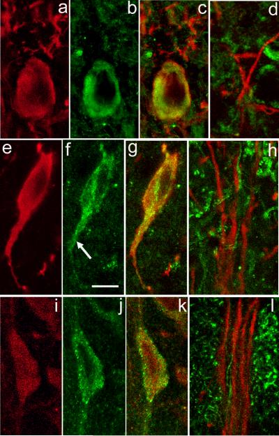
Figure 1. ER Ca2+ store protein immunoreactivity in nigral DAergic neurons.
a-c. SERCA2 immunoreactivity in DAergic neurons in the SNc. Immunostaining for TH (red) (a) and SERCA2 (green) (b) with merged images for TH and SERCA (overlap appears yellow) (c). In the perikaryal cytoplasm, SERCA2 immunoreactivity is observed as a ring around the nucleus that extends almost to the perikaryal edge. Additional SERCA staining is seen within the most proximal portion of TH-ir dendrites (TH-ir profiles above perikaryon in c), but is absent from DAergic dendrites within SNr (d). e-g. Localization of IP3Rs in DAergic neurons in the SNc. Immunostaining for TH (red) (e) and IP3R (green) (f) with merged images for TH and IP3R (g). Note that immunoreactivity to IP3R and TH colocalize in the perikaryon and extends down a proximal dendrite (f, arrow). h. In SNr, however, IP3R immunoreactivity (green) does not colocalize with that of TH (red), implying minimal IP3R expression in distal DAergic dendrites. i-k, Localization of RyRs in DAergic neurons in the SNc. Immunostaining for TH (red) (i) and RyR (green) (j) with merged images for TH and RyR (k). Note that RyR immunostaining includes a mixture of large puncta located at the edge of the perikaryon and smaller puncta located in the perikaryal cytoplasm. RyR immunoreactivity (green) does not colocalize with that of TH within SNr (l), implying low levels in distal dendrites. Scale bar in panel f is 10 μm and applies to all panels.

