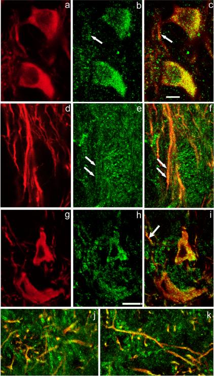
Figure 2. mGluR1 and Cav1.3 immunoreactivity in nigral DAergic neurons.
Immunostaining for TH (red) (a) and mGluR1α (green) (b) with merged images for TH and mGluR1α (overlap appears yellow) (c). The mGluR1α immunoreactivity appears as puncta that colocalize with TH in the perikarya and primary dendrites of DAergic neurons in SNc. Arrows in (b) and (c) indicate corresponding locations at which colocalization of mGluR1α- and TH-ir occurs in dendritic processes within SNc. d-f. A moderate level of mGluR1α staining is seen within TH-ir dendrites in SNr. Paired arrows in (e) and (f) point to colocalization of mGluR- and TH-ir in a dendritic profile. g-i. Localization of Cav1.3, an L-type Ca2+ channel subunit, in DAergic neuronal perikarya in the SNc. Immunostaining for TH (red) (g) and Cav1.3 (green) (h) with merged images for TH and Cav1.3 (i). Cav1.3 immunoreactivity is punctate. j-k. Colocalization of immunoreactivity to TH and Cav1.3 in DAergic processes at the border of SNc/SNr (j) and deep within SNr (k). Scale bar in panel c is 10 μm and applies to all panels.

