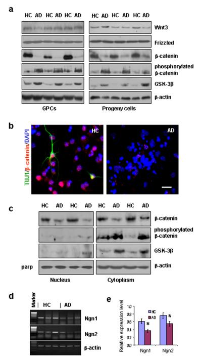
Figure 3.

β-catenin signaling and proneural gene transcription in AD vs. HC GPCs and their progeny. (a). Western blot showed a reduction of non-phosphorylated β-catenin levels and an increase in both phosphorylated β-catenin and GSK-3β levels in AD GPCs and their progeny compared to their HC counterparts, without significant differences in either Wnt3 or frizzled expression. (b). Immunostaining for βIII-tubulin (mature neurons), β-catenin, and DAPI (all nuclei) showed a decrease in β-catenin expression in AD differentiating cells compared to HC cells. (c). Western blot showed a decrease in non-phosphorylated β-catenin expression and in the nuclear fractions of AD GPC progeny compared to HC progeny with no detectable GSK-3β or phosphorylated β-catenin expression in AD or HC progeny. The non-phosphorylated β-catenin levels also decreased in the cytoplasmic fraction of AD progeny compared to HC progeny; however, this change was accompanied with increased levels of GSK-3β and phosphorylated β-catenin. (d). RT-PCR revealed that mRNA levels of Ngn1 and Ngn2 were reduced in AD GPC progeny compared to HC progeny. (e). Spot densometric analysis by Fluchem8900 software was used to quantify the RT-PCR relative expression levels of Ngn1 and Ngn2 mRNA in the AD and HC GPC progeny. The expression levels were normalized to the respective β-actin levels (Student’s t test, *p < 0.05, n = 3 per group).
