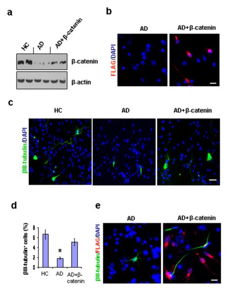
Figure 4.

Effect of β-catenin transfection on neurogenesis in AD GPC progeny. Single passage NG2+ cells from AD brains were virally transfected with either β-catenin cDNA or a control vector for 5 hrs, and the culture was extended to 14 days. The transfected AD, control AD, and control HC neurospheres were dissociated into single cells and were cultured in differentiation medium for another 4 days. (a). Western blot revealed that the expression level of β-catenin in the transfected AD GPC progeny was significantly greater than in the control AD progeny but less than in the control HC progeny. (b). The success of the β-catenin transfection was confirmed by immunostaining the transfected and control AD GPC progeny with DAPI and an anti-FLAG-tag antibody. Approximately 40% of the DAPI-labeled cells in the transfected AD GPC progeny were FLAG+, while no control AD progeny were FLAG+. (c). Among the control HC, control AD, and β-catenin transfected AD GCP progeny, the neurons were visualized by βIII tubulin staining (green), and the nuclei were counterstained with DAPI (blue) (n = 2 per group). (d). Immunoreactive quantification among the control HC, control AD, and transfected AD progeny revealed that the percentage of mature neurons among the β-catenin transfected AD GPC progeny was significantly restored(n = 3 per group, ANOVA, *p < 0.05). (e). The β-catenin transfected AD GPCs produced more new neurons (βIII-tubulin+) than did the control AD GPCs. Among the transfected AD GPC progeny, approximately 70% of the new neurons developed from virally infected AD GPCs (indicated by the co-localization of βIII-tubulin and and FLAG-tag immunostaining). Scale bars: 50 μm (c), 30 μm (b), (e).
