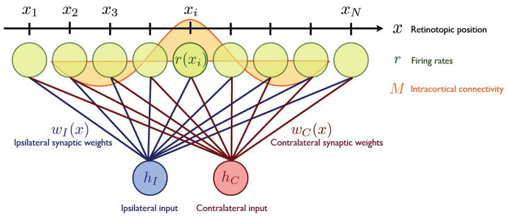
Figure 1.
A schematic figure of the model. N cortical neurons are lined up on a one dimensional axis x. Each neuron receives feedforward input from both ipsilateral and contralateral eyes and those synapses are modified according to activity dependent plasticity rules. The intracortical connectivity, M, is a function of the distance between two cortical positions and changes its profile at the onset of the critical period when cortical inhibition matures.

