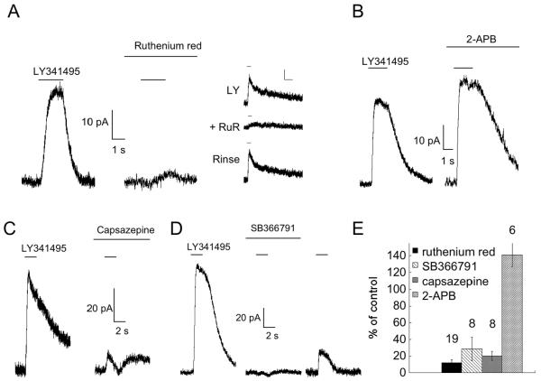
Fig. 1. The rod bipolar cell transduction current is blocked by antagonists of TRPV1.
A. Response to 100 μM LY341495 (delivered via fast flow apparatus) before (left panel) and after a 5 minute application of 10 μM ruthenium red (center panel). Right panel, response of another cell to a 1 second puff of LY341495 delivered through a puffer pipet alone (top), or during simultaneous application of ruthenium red from a second puffer pipet (middle). Ruthenium red was applied alone for 10 seconds prior to obtaining the middle trace. The inhibition of ruthenium red was readily reversed using this approach (bottom). Scale bars: 10 pA and 2 seconds. Right panel is from a different cell. B. Response to LY341495 before and after a 5 minute application of 100 μM 2-APB. C,D. Responses to 1 second puffs of LY341495 (100 μM) before and after 5 minute bath application of 20 μM capsazepine (C) and 20 μM SB366791 (D). Responses to LY341495 typically showed partial recovery after removal of antagonists, as shown in the right panel of (D). Traces in (C) and (D) are from different cells. E. Summary of results. The number of cells for each experiment is indicated above each bar.

