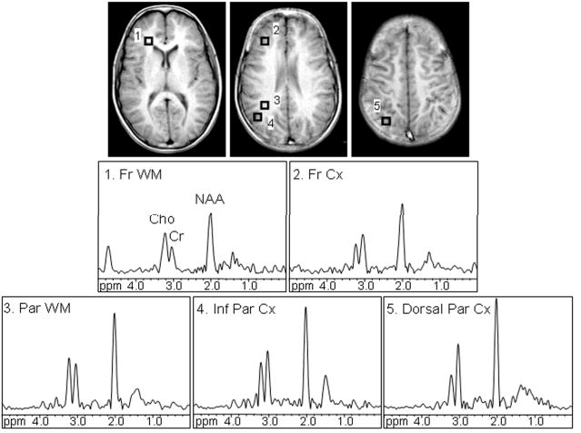
FIGURE 1.

Axial T1-weighted spin-echo images display regions of interest in the frontal white matter (1, Fr WM), dorsolateral prefrontal cortex (2, Fr Cx), parietal white matter (3, Par WM), inferior parietal cortex (4, Inf Par Cx), and dorsal parietal cortex (5, Dorsal Par Cx). Proton MR spectra corresponding to the evaluated regions of interest are shown. Signals of choline (Cho), creatine (Cr), and N-acetyl aspartate (NAA) were detected. All spectra are equally scaled.
