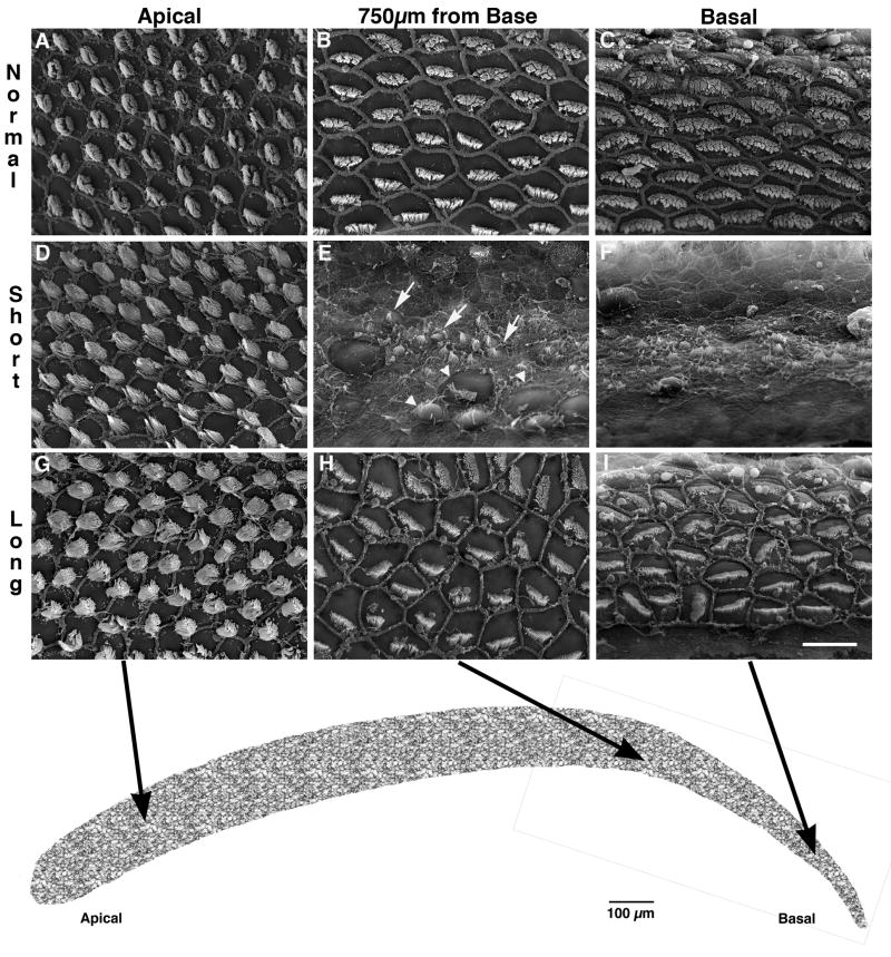
Fig. 5.
Higher magnification scanning electron micrographs of normal (A-C), lesion – short recovery (D-F), and lesion – long recovery (G-I) starling basilar papillae shown in Fig. 4. The low-frequency (apical) region of all cases is unaffected by the kanamycin paradigm (A, D, G). Immature, regenerating hair cells (arrows) can be seen adjacent to remaining, damaged hair cells (arrowheads) in the region 750 μm from the basal tip (E) and in the basal end (F) of the BP from the short-recovery animal. This morphology is in stark contrast to that seen in the normal starling in the same regions (B and C). At longer survival times, regenerated hair cells attain a more normal appearance, although stereocilia bundles are not uniformly oriented and may display abnormal bundle morphology (H and I). The schematic representation of the entire starling basilar papilla at the bottom shows the regions from which the higher magnification photomicrographs were taken. Bar in I represents 10 μm in A-I.

