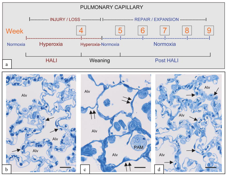
Figure 1. In vivo model of capillary remodeling in HALI and post-HALI.
Schema of in vivo model (a) and representative brightfield images illustrating patent capillaries in normal lung (b, arrows), their loss in HALI (c, arrows, week 4), and the restoration of patent capillaries early in the post-HALI phase following spontaneous repair (d, arrow, week 6). In the model rodents breathe 87% oxygen for 4 weeks (HALI), or 87% oxygen for 4 weeks, followed by weaning to air for 1 week (week 5) and air breathing for up to 4 weeks (post-HALI, weeks 6-9). Initially in HALI (week1), endothelial cells of small vessels and capillaries in the alveolar-capillary membrane are severely injured. As these cells (and epithelial cells) adapt to the high oxygen tension, the membrane remodels (HALI, weeks 2-4); however, there is extensive capillary loss. Because of the greatly reduced capillary bed present at this time, the oxygen tension is lowered (∼less 10%) daily during the transition to breathing air (week 5) to prevent asphyxia, dyspnea and cyanosis – the lower oxygen tension of air (i.e., relative hypoxia) triggering a burst of spontaneous vascular repair post-HALI (weeks 6 and 7). Spontaneous capillary repair and expansion continue to restore the membrane post-HALI until, as patent capillary networks approach their normal distribution, the response wanes (week 9) [8]. 2μm-thick resin sections stained with toluidine blue. Bars = 25μm (b-d).

