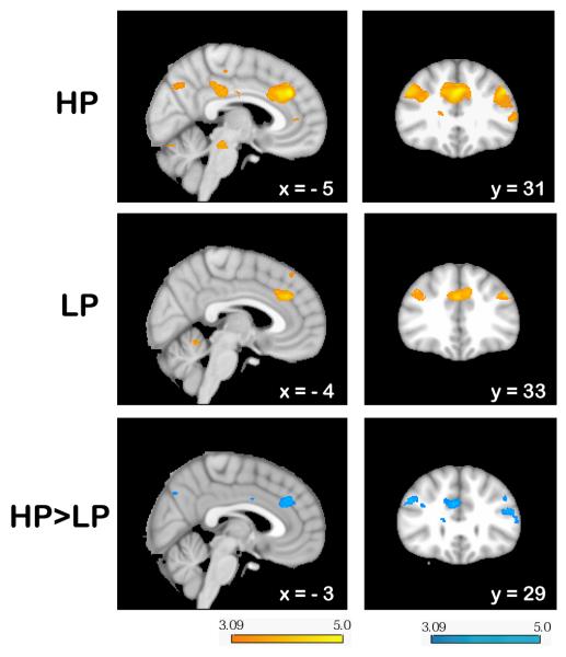
Fig. 4.
Signal change in response to reversal errors (REVERR — ALLPOS) superimposed on the MNI template brain. In both conditions (HP: top row; LP: middle row), there was increased activity in the RCZ (left) and in the lateral prefrontal cortex (right). Both the response of the RCZ and the lateral prefrontal cortex were more pronounced in the HP than in the LP condition. The color bars indicate z-scores. See Table S2 for a comprehensive list of all activations.

