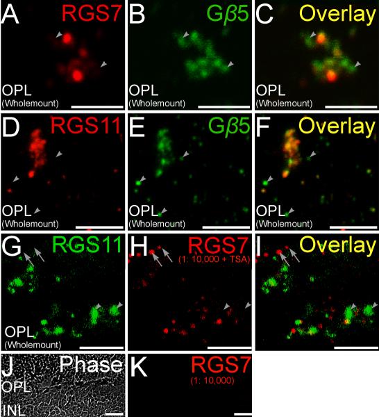
Figure 4. Double staining for RGS proteins and Gβ5.
Double labeling using antibodies directed against RGS7 and RGS11 (red) and Gβ5 (green). A-C (whole-mount), A magnified wholemount view of the OPL shows ON-cone bipolar cells in tight clusters. Some of the Gβ5 immunopositive puncta did not have significant labeling for RGS7 (arrowheads). D-F (whole-mount), RGS11 immunopositive puncta in the OPL showed good co-localization with closely spaced clusters of Gβ5 immunopositive puncta belonging to ON cone bipolar cells. However, isolated Gβ5 immunopositive puncta likely belonging to rod bipolar cells did not have detectable levels of RGS11 immunofluorescence (arrowheads). G-I, Non-coincident immunofluorescence for RGS7 or RGS11 in the OPL (whole-mount). Double labeling using antibodies directed against RGS11 (green) and RGS7 (red). Staining for RGS7 and RGS11 did not overlap for some dendritic tips (arrowhead, RGS11+, RGS7−; arrow, RGS7+, RGS11−). Scale bar = 5μm, A-I. J, K(radial section), Tyramide signal amplification (TSA) did not produce any visible immunolabeling at the dilution of 1:10,000 for the red channel, eliminating the possibility of cross reaction of the secondary antibodies in the red channel with the primary in the green channel. Scale bar = 10μm (J, K).

