Fig. 1.
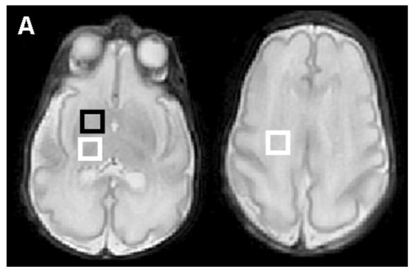
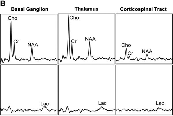
Location of the voxels and normal neonatal proton spectra obtained at gestation age of 32 weeks from a prematurely born infant. The infant received pentobarbital sedation. (A) The basal ganglion voxel includes the head of caudate and the anterior putamen (black box, left image). The thalamic voxel is indicated by the white box in the left image. The corticospinal tract voxel includes the corticospinal tract within the centrum semiovale (white box in the right image). (B) Lactate-edited spectra. Top row shows the summed spectra in each voxel for Cho, Cr, NAA. The bottom row shows the difference spectra for each voxel; the difference spectra show only the Lac peaks. Cho, choline; Cr, creatine; NAA, N-acetylaspartate; Lac, lactate.


