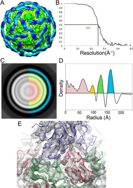
Figure 7. Analysis of three-dimensional reconstruction of DAV particles.
A. Surface representation of the DaV CryoEM reconstruction contoured at 2 sigma. The inner N-terminal domain (green) is well ordered with visible secondary structural features, while the outer C-terminal domain (blue) contains only very low-resolution features. B. The estimated resolution of the reconstruction is 8Å according to FSC criteria at 0.5. C. A radially averaged slice through center of reconstruction. Blue = C-terminal domain, Green = N-terminal domain, Orange, Red = RNA D. Plot showing density by radius (colours similar to C.) E. Docking of the beta-sandwich domain of the TBSV crystal structure into the N-terminal domain of the DaV cryoEM density.

