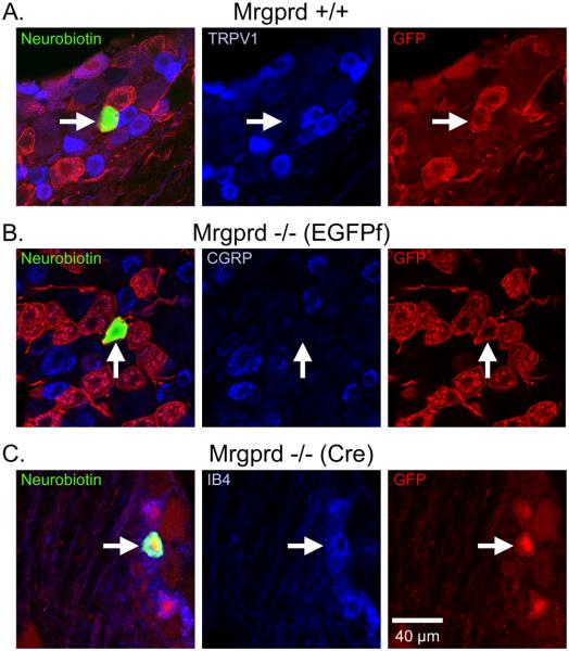
Figure 1. CPMs in Mrgprd knockin and WT mice bind IB4, but do not express either TRPV1 or CGRP.
Sample immunohistochemistry of recorded CPM cells in (A) Mrgpr+/+, (B) Mrgprd−/− (EGFPf ) and (C) Mrgprd−/− (Cre) as indicated by biotin-labeling (left panels, green). Labeling of TRPV1, CGRP and IB4 are shown for Mrgprd-IRES-EGFPf+/+, Mrgprd−/− (EGFPf) and Mrgprd−/− (Cre), respectively (middle panels, blue). GFP labeling is shown in the right panels (red). Arrows indicate recorded cell. Scale bar represents 40 μm (C, right panel).

