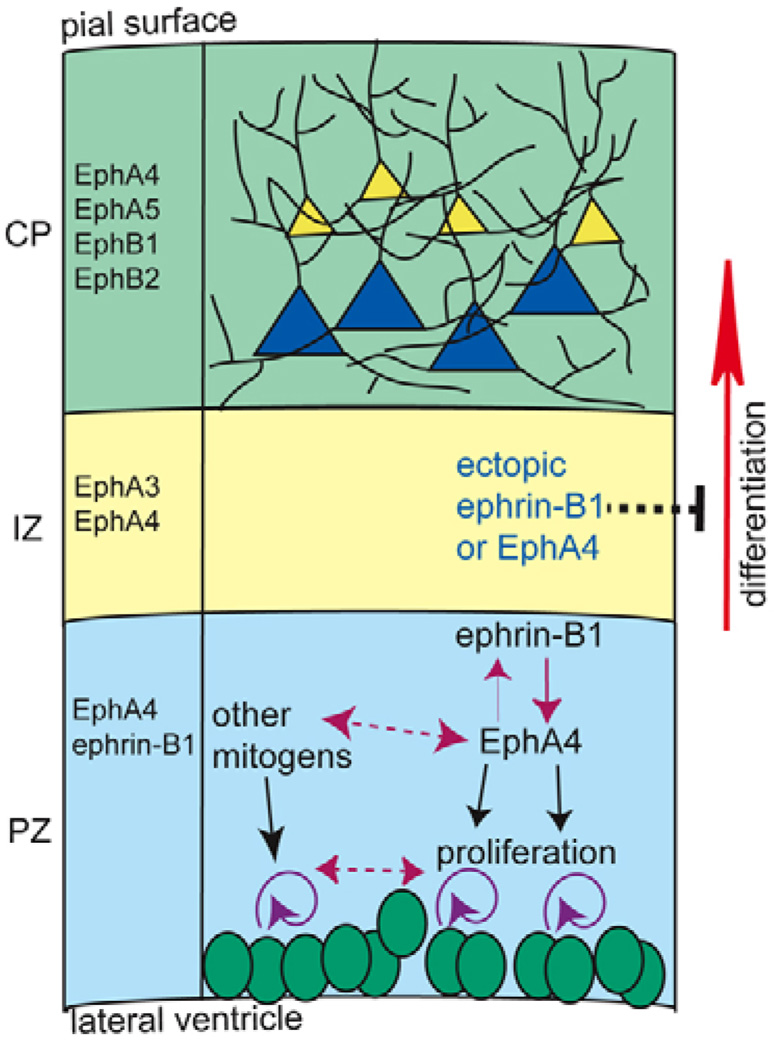
Fig. 8. Schematic of Eph signaling within the cerebral wall.
In the proliferative zones (PZ), ephrin B1 and EphA4 are expressed (light blue) and ephrin B1-EphA4 signaling (pink solid arrow) promotes cell proliferation (purple circles) in balance with other mitogens (pink dashed arrows). Embryonic zones containing postmitotic neurons, the intermediate zone (IZ) and cortical plate (CP), express unique subsets of Eph family members (yellow and green, respectively). Ectopic expression of ephrin B1 in postmitotic populations or EphA4 in progenitors antagonizes (black dashed inhibitory bar) neuronal differentiation (red arrow).

