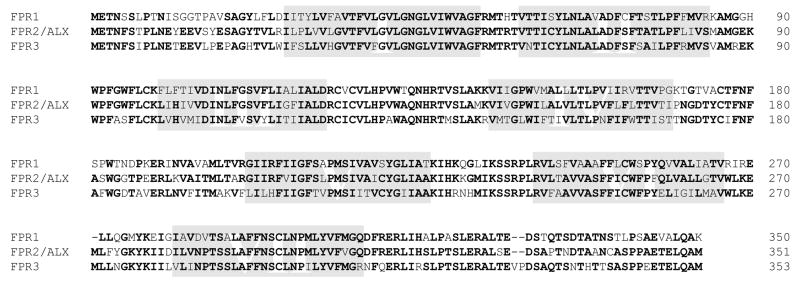
Figure 2. Alignment of the protein sequences of the human FPRs.
The putative transmembrane domains (TM-I to -VII) are highlighted in shades. Sequence of the FPR-98 isoform is shown. Comparison of the three receptors has identified highly conserved regions including most of TM-I and TM-II, and the short intracellular loop connecting TM-I and TM-II. The second intracellular loops from these receptors, known for G protein interaction, are nearly identical. TM-VII, including the NPXXY motif and a stretch of ~25 amino acids extending towards the C-terminal tail, are also conserved among these receptors. Major differences are found in the extracellular domains between FPR1 and the other two receptors, especially in the amino termini (~50% different), the second extracellular loops (56% different), and the third extracellular loop (~50% different). The two putative N-glycosylation sites in the N-terminal domains are conserved among all three receptors. Most of the serines and threonines in the C-terminal tail, along with charged residues that constitute consensus GRK phosphorylation sites, are also conserved among these receptors.

