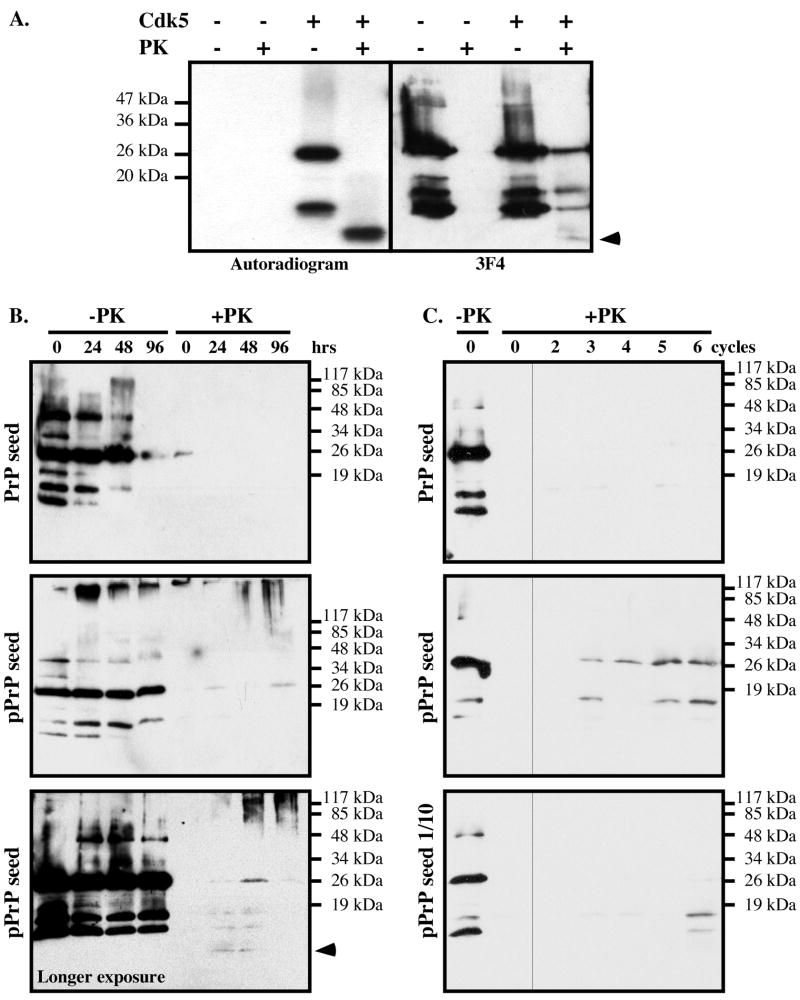
Figure 3. Cdk5-phosphorylated PrP induces the aggregation of non-phosphorylated PrP in vitro.
A. Autoradiogram and western blot analysis with 3F4 antibody of non-phosphorylated or Cdk5-phosphorylated PrP treated with 10 μg/mL PK for 1 hr at 37°C. The arrow indicates the 10 kDa PrPRES fragment detected on the autoradiogram or western blot. B. PrP western blot of non-phosphorylated PrP seeded with kinase assays performed with (pPrP) or without Cdk5, and incubated for the indicated time without (-PK) or with PK treatment (+PK). The lower panel shows a longer exposure of another pPrP seeded experiment revealing the 10 kDa PrP fragment in +PK. C. PrP western blot of non-phosphorylated PrP seeded with a 2 μl aliquot of the 96 hr time point (0 cycle) in 3B and incubated 24 hrs (cycle 1). Subsequent cycles represent samples where 2 μl at the end of the incubation period was added into fresh non-phosphorylated PrP and incubated 24 hrs. The lower panel represents an original seed of 0.2 μl of the 96 hr time point in B.

