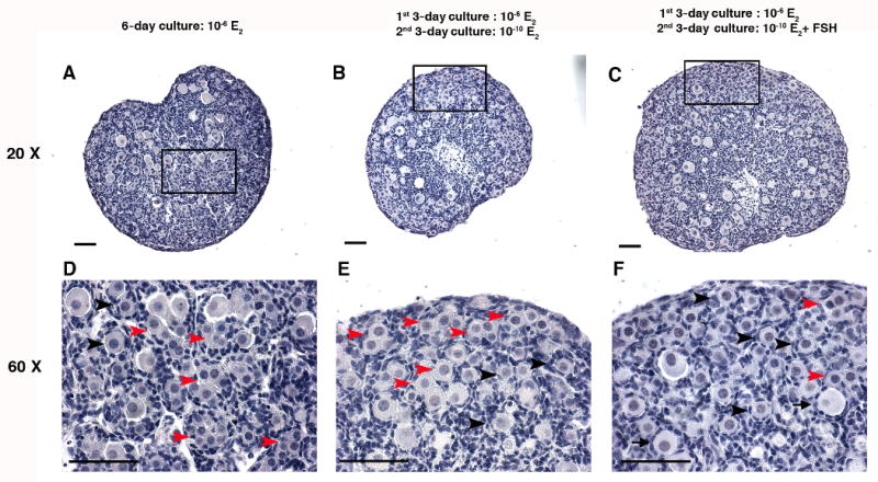
Figure 2. The histology of the primordial folliculogenesis in in vitro cultured 17.5 dpc mouse fetal ovaries.

Histology of 17.5 dpc mouse fetal ovaries cultured in vitro in (A) 10−6 M E2 for 6 days; (B) 10-6 M E2 for 1st 3 days then 10-10 M E2 for 2nd 3 days; or (C) 10-6 M E2 for 1st 3 days and 10-10 M E2 plus 10 mIU/ml FSH for 2nd 3 days. (D-F) Higher magnification shows the structure of the unassembled oocytes, primordial follicles and primary follicles and in the ovaries cultured under the three conditions listed in A-C. Red arrowheads indicate unassembled oocytes: oocytes that did not surround by pre-granulosa cells completely or oocytes were associated; black arrowheads indicate primordial follicles: one oocyte was surrounded by several flatten pre-granulosa cells; black arrows indicate primary follicles: one oocyte with expanded cytoplasm surrounded by a complete layer of cuboidal granulosa cells. Bar=50 μm.
