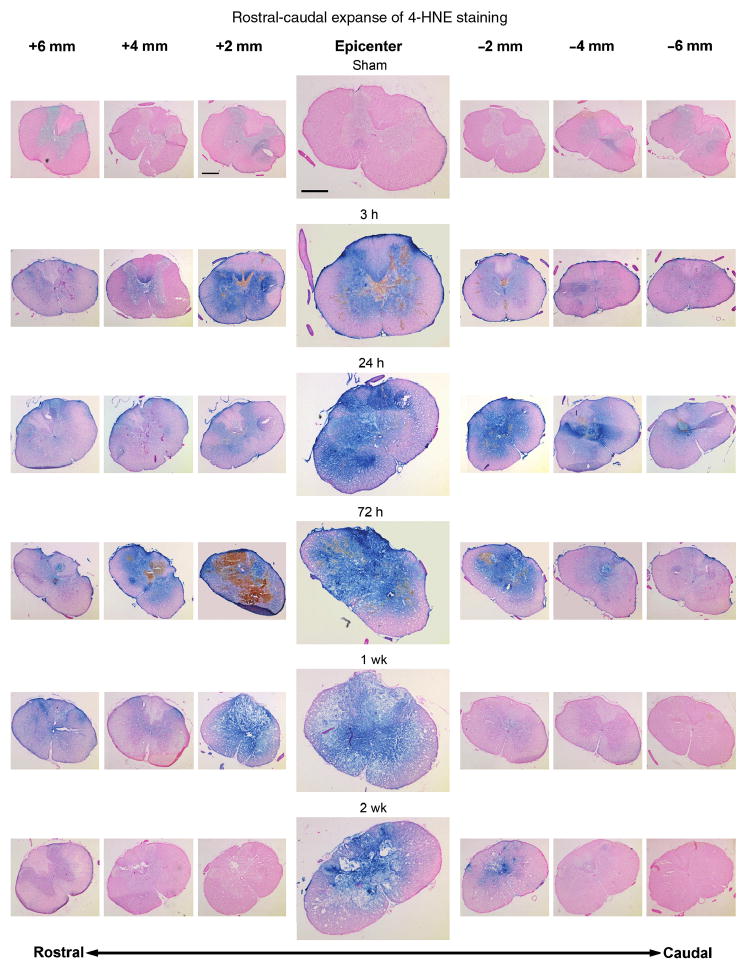
FIG. 2.
Rostral-caudal extent of 4-HNE staining at all time-points as shown by immunohistochemistry. All sections were counterstained with nuclear fast red. Images were taken every 2 mm starting 6 mm rostral to the epicenter and ending 6 mm caudal to the epicenter. At each time point, the images were taken from only one animal. By 3 h post-injury, staining was elevated above sham levels at the epicenter, with this elevation present at least 6 mm in both the rostral and caudal directions. This staining pattern persisted up to 72 h post-injury, with 4-HNE indicated at least as much as 6 mm from the epicenter in both directions. At 1 week post-injury, staining was still intense 2 mm rostral to the epicenter, but had abated somewhat by 4 mm, although it remained above sham levels out to 6 mm rostrally. Very little staining was seen at 1 week post-injury in the caudal direction. By 2 weeks post-injury, staining was still present at the epicenter, but was not seen more than 2 mm in either direction. Compare Figure 2 with Figure 3, which shows protein nitration-related 3-NT staining of the adjacent sections (scale bar =500 μm).

