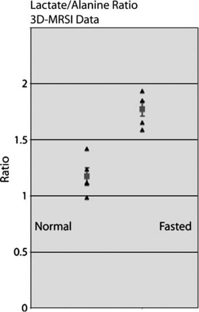
Fig. 6.
Lactate area to alanine area from 3D-MRSI studies (averaged over liver voxels per rat) of normal and fasted liver. Normal rat liver showed a significantly lower lactate area to alanine area ratio (P<0.01) than fasted rat liver. These data corroborate the dynamic MRS data, with the means matching closely and indicating the same conclusion. Mean/SD—(normal 1.18±0.16, fasted 1.78±0.15). (Note: triangular markers show the collected data points and the square marker/error bars show the mean/standard errors.)

