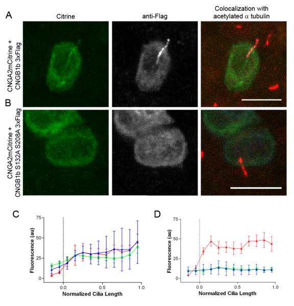
Figure 3. Mutation of CK2 Phosphorylation Sites on the N-terminus of CNGB1b Impairs Ciliary Trafficking of the CNG Channel.

Representative confocal images of MDCK cells transfected with CNGA2-mCitrine and (A) CNGB1b-3xFlag or (B) CNGB1b S132A, S208A-3xFlag. Citrine signal is on left (green). Flag immunostaining for CNGB1b is in middle (grayscale). Merged image with staining for acetylated α tubulin (red) is on right. Bar represents 10 μm. Average data from multiple cells (n=6-7) shown for (C) CNGB1b wild-type or (D) CNGB1b S132A, S208A. Data were apportioned into bins of 1/10th of normalized cilia length. Acetylated tubulin signal shown in red, CNGA2-mCitrine fluorescence shown in green, and anti-Flag immunostaining signal shown in blue (arbitrary fluorescent units).
