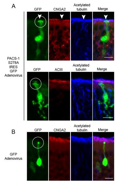
Figure 7. Expression of Mutant PACS-1 in Native OSNs Causes Mislocalization of the CNG Channel, but not ACIII.
Representative collapsed confocal images of coronal sections of OE from adenovirally-infected mice. (A) (top) Immunostaining of a PACS-1 S278A IRES GFP-infected OSN (green) with antibodies against CNGA2 (red) and acetylated α tubulin (blue) demonstrates a loss of ciliary CNG channel from the infected OSN (white arrowhead) with no change in the cilia layer. (bottom) Immunostaining of a PACS-1 S278A IRES GFP-infected OSN (green) with antibodies against ACIII (red) and acetylated α tubulin (blue) demonstrates no detectable change in ciliary ACIII or the cilia layer from the infected OSN. (B) Immunostaining of a GFP-infected OSN (green) with antibodies against CNGA2 (red) and acetylated α tubulin (blue) demonstrates no detectable change in ciliary CNG channel or the cilia layer from the infected OSN. For all conditions, merged image shown on right (Merge), white circles mark dendritic knobs, and bars represent 10 μm.

