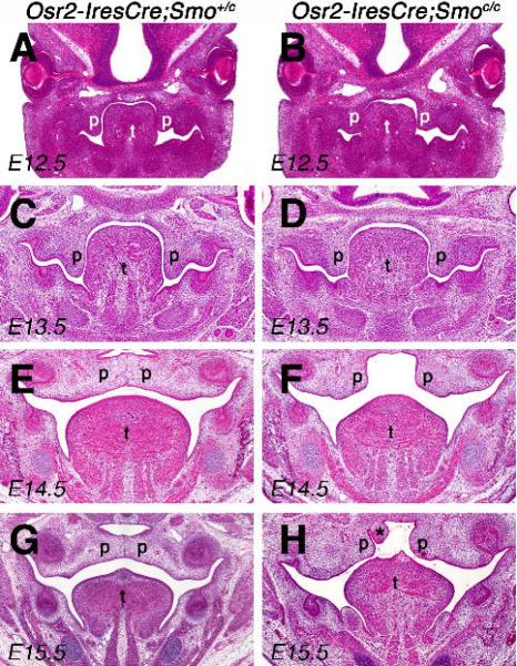
Fig. 4.
Histological analyses of palate development in the control and Osr2-IresCre;Smoc/c mutant embryos. (A, B) HE-stained frontal sections of E12.5 Osr2-IresCre;Smo+/c (A) and Osr2-IresCre;Smoc/c mutant (B) embryos showed comparable initial outgrowth of palatal shelves. (C, D) At E13.5, the palatal shelves of Osr2-IresCre;Smoc/c mutant embryo (D) appeared slightly retarded distally in comparison with the Osr2-IresCre;Smo+/c embryo (C). (E, F) At E14.5, while the palatal shelves had elevated and initiated fusion at the midline in the control embryo (E), the palatal shelves of the Osr2-IresCre;Smoc/c mutant embryo (F) appeared severely retarded and failed to contact each other. (G, H) Some Osr2-IresCre;Smoc/c mutant embryos exhibited tissue protrusion into the nasopharynx, shown in H (marked by an asterisk), which was never observed in control embryos. p, palatal shelf; t, tongue.

