Fig. 2.
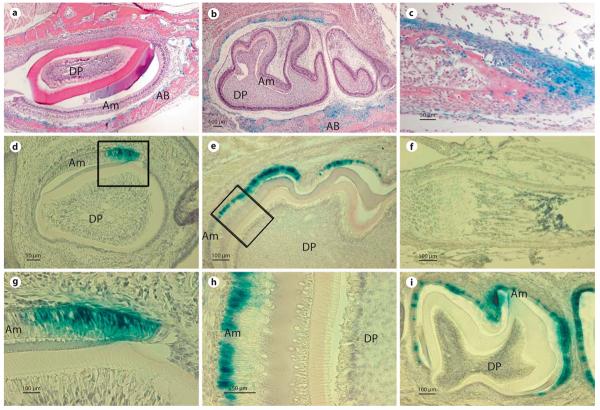
Comparison of β-galactosidase activity expressed by the 3.9- and 5.2-kb Enam-LacZ transgenes. a, b β-Galactosidase expression was not detected in ameloblasts (Am) or dental pulp (DP) cells of day 1 incisors (a) or day 1 and 5 molars (b). In contrast, β-galactosidase activity was detected in alveolar bone (AB) surrounding the incisors and molars. c At postnatal day 1, staining of long bones of the extremities is also distinct; mostly from the cells that encapsulate the developing long bones and joints. d–i β-Galactosidase activity was detected in ameloblasts (Am) of postnatal day 1 incisor (d, g). In molars, β-galactosidase reporter gene activity was not found in day 1 molars and it was first detected in the ameloblasts at postnatal day 5 (e, h). Positive staining of β-galactosidase activity specifically localized to the nuclei of the ameloblasts (g, h). The intensity of β-galactosidase activity peaked at postnatal day 9 and continued in maturation stage ameloblasts, being located within the nuclei until postnatal day 17 (i). Dental pulp (DP) and alveolar bone (AB) cells were devoid of staining. No β-galactosidase activity was detected in long bones of postnatal day 12 mice (f).

