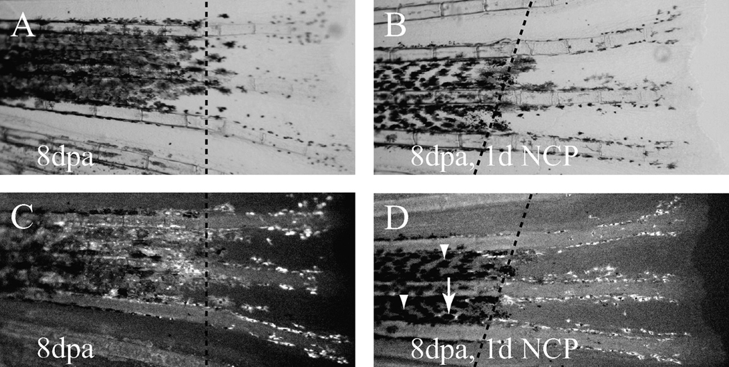
FIG. 7.

NCP does not ablate newly regenerated melanocytes. (A) Fish fin carrying transgenic tyrp1:eGFP 8 days postamputation (dpa) under white light. The dashed line represents the amputation plane. (B) A fish fin carrying transgenic tyrp1:eGFP 8 dpa and 1 day of NCP treatment under white light. The dashed line is the amputation plane. (C) The same fin as in (A), under fluorescent light. Note GFP is on both sides of the amputation plane in all melanocytes. (D) The same fin as (B), under fluorescent light. Most of the melanocytes proximal (to the left) of the amputation plane have lost GFP expression (arrowheads), although some retain GFP expression (arrow). All of the melanocytes distal (to the right) of the amputation plane express GFP.
