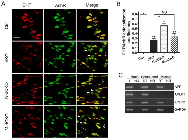
Figure 3.
Aberrant CHT localization in presynaptic terminals of APP mutant NMJ. A. Whole mount diaphragm muscles from P0 control (Ctrl), dKO, N-dCKO or M-dCKO animals were stained with anti-CHT antibody or α-BTX (AchR). The images were captured by confocal microscopy and displayed either as individual staining or merged (last row). Representative endplates with poor CHT coverage were marked by arrowheads and extra-synaptic CHT staining by arrows. Scale bar: 20 μM. B. Quantification of the percentage of AchR-positive endplates covered by CHT (average ± SEM of 20 endplates/genotype). Asterisks above each bar are in comparison with the control. Asterisks on top of the brackets are in comparison with the dKO mice. *p<0.05, **p<0.01, NS: non-significant (p>0.05) (one way ANOVA). C. RT-PCR analysis of APP, APLP1 and APLP2 expression in P0 brain, spinal cord and muscle samples. RT: with reverse transcriptase; NR: the same sample and reaction in the absence of reverse transcriptase. GAPDH was used as amplification control.

