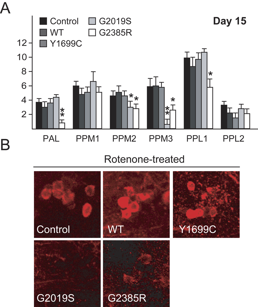
Figure 3. Exposure to rotenone accelerates DA degeneration in LRRK2 G2019 and G2385R mutant flies.
(A) Bar-graph showing the number of TH-positive DA neurons in different clusters of various fly species at 15-day after rotenone treatment (n=15). (B) Representative confocal microscopy images showing TH-positive (red) DA neurons in the PPM3 cluster of various, rotenone-treated, fly species, as indicated. (Genotype: ddc-Gal4/+ or ddc-Gal4-hLRRK2)

