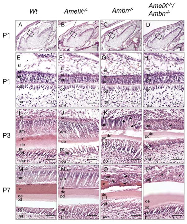
Fig. 2.
Histological analysis of teeth from the wild-type, Amel X −/−, Ambn−/−, and Amel X−/−/Ambn−/− mice. Hematoxylin-eosin staining of the sagittal sections of the mandibular first molars of P1 (A–H), P3 (I–L), and P7 (M–P) wild-type and mutant mice: wild-type (A, E, I and M), Amel X−/− (B, F, J and N), Ambn−/− (C, G, K and O) and Amel X−/−/Ambn−/− mice (D, H, L and P). P3 and P7 Ambn−/− ameloblasts display multiple layers containing abnormal calcified structures (Fig. 2K and 2O, arrows). Amel X−/−/Ambn−/− ameloblasts also form multiple layers, however, they do not contain the calcified structures (Fig. 2L and 2P, arrowhead). am, ameloblast; si, stratum intermedium; e, enamel; pd, predentin; de, dentin; od, odontoblast; pu, pulp sr, stellate reticulum). Bars in A–D = 500 μm; bars in E–P = 50 μm.

