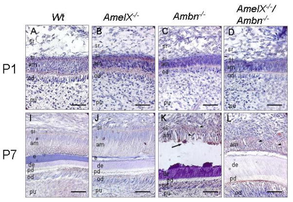
Fig. 3.
RhoGDI expression in the ameloblasts of the wild-type, Amel X−/−, Ambn−/−, and Amel X−/−/Ambn−/− mice. Sagittal sections of the incisors from the wild-type (A and E), Amel X−/−(B and F), Ambn−/− (C and G), and Amel X−/−/Ambn−/− mice (D and H) were stained with the RhoGDI antibody as described in Materials and Methods. Note positive staining in the detached ameloblasts of Ambn−/− (C and G) and Amel X−/−/Ambn−/− mice (D and H). Bars = 50 μm. am; ameloblast, od; odontoblast, pu; pulp, si, stratum intermedia, e; enamel, d; dentin, pd, predentin

