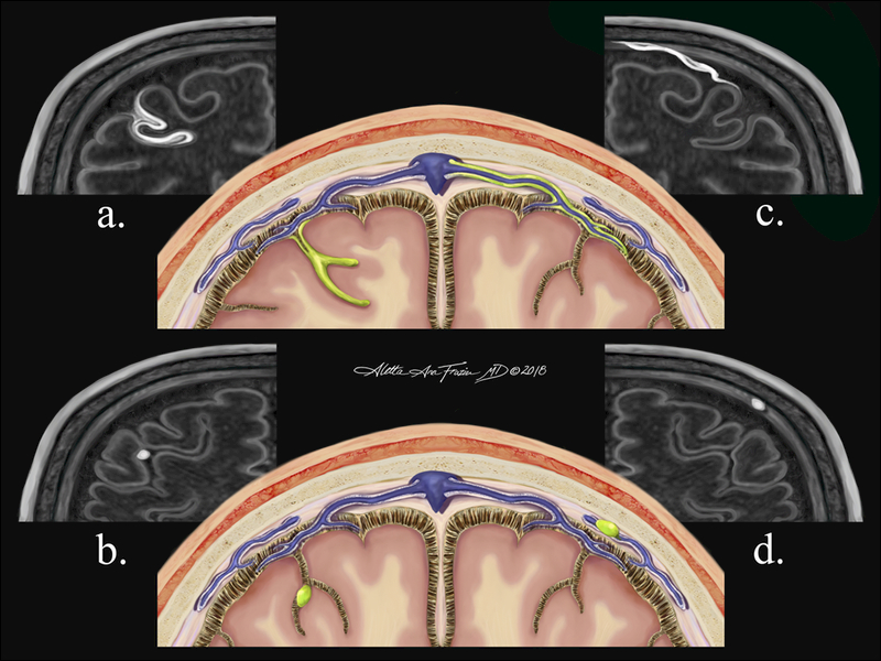
Figure 1:

Original illustration depicting the four morphologies of meningeal enhancement seen in this analysis. Subarachnoid spread/fill pattern (represented by green coloring in a) is an amorphous and ill-defined collection of contrast pooling within cerebral sulci. Subarachnoid nodular pattern (b) is defined as a punctate, discrete site of meningeal enhancement located within cerebral sulci abutting the pial surface. Venous rim pattern (c) is characterized by extension of contrast along the outer margin of large meningeal vessels with preserved internal flow void creating a characteristic tram-track appearance. Dural nodular pattern (d) is a circumscribed, rounded focus of contrast situated along the dural margin without extension into the subarachnoid space. The perivascular, tubular white structures (seen in schematics a,b,&d) represent the recently discovered meningeal lymphatic system. Reaccumulation of leaked contrast from the cerebrospinal fluid into these meningeal lymph channels is a potential mechanism for the venous rim pattern (c).
