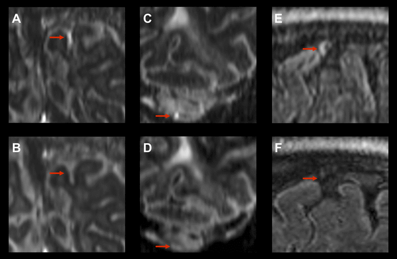
Figure 4:

Examples of resolving foci of meningeal enhancement on delayed post-contrast FLAIR at 7T. Coronal reformatted images show subarachnoid spread/fill enhancement that resolves between October 23, 2014 (A) and February 26, 2016 (B) in a 49 year-old woman with relapsing-remitting MS. Coronal reformatted images show subarachnoid nodular enhancement within cerebellar folia that resolves between October 8, 2014 (C) and February 19, 2016 (D) in a 49 year-old man with relapsing remitting MS. Sagittal formatted images show vessel wall enhancement that resolves from May 9, 2016 (E) to May 31, 2017 (F) in a 44 year-old woman with secondary progressive MS. Note that no foci of meningeal enhancement classified as dural subtype resolved in this study.
