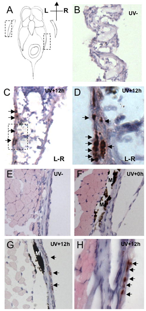
Figure 4.

Visualization of DNA damage sites in zebrafish fin (B-D) and skin (E-H). Histological sections of wild type and p53 mutant zebrafish 12 hours after of UVB (2.16 kJ/m2) exposure were stained with phospho-histone H2AX antibody. A. Schematic diagram of zebrafish section to denote fin and skin tissues. B. Section of pectoral fin of untreated wild type fish, magnification 200× C. Section of pectoral fin of UVB exposed wild type fish. Signals are located in the epidermis on the most outer side of fin. Stained cells are indicated with arrows, 200× D. High magnification (630×) view on to the stained nucleus in epidermis in the fin, as outlined in the boxed region of C. E. No staining is detected in the skin of untreated fish, magnification 200× F. Skin of fish sacrificed immediately after UVB exposure reveals no staining for phospho-H2AX, magnification 200×. G. Skin at 12 hours post irradiation reveals specific phospho-H2AX staining, magnification 200× H. High magnification (630×) view on to the stained nucleus in the skin. Phospho-H2AX positive nuclei are indicated with arrows; M denotes melanin.
