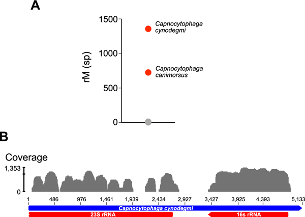
Figure 2: Identification of C. canimorsus and C. cynodegmi by metagenomic deep sequencing.

(A) Organisms identified in the patient’s aqueous sample are plotted as a function of matched read pairs per million read pairs (rM) at the species level based on nucleotide alignment. Sequencing reads aligned to C. canimorsus and C. cynodegmi (red circles) predominated the sample. Grey circles indicate background sequencing reads. (B) C. cynodegmi sequences from Case 2 assembled against the reference C. cynodegmi genome (GenBank NZ_CP022378).
