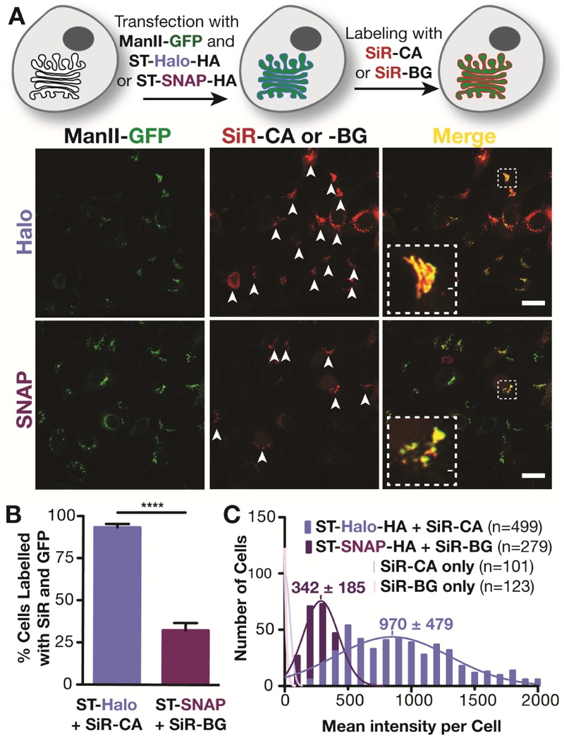
Figure 1.

Comparison of Golgi labeling with Halo- and SNAP fusion proteins of sialyl transferase. A) Top: Scheme of the labeling procedure; Bottom: Confocal images of live HeLa cells that have been treated as described in the scheme above. The white arrows indicate cells that express ManII-GFP and have been labeled with SiR-CA or SiR-BG. Scale bar: 20 μm. B) Quantification of cells expressing ManII-GFP that are positive for SiR from three independent experiments (ST-Halo: 740 cells in total; ST-SNAP: 837 cells in total). C) Fluorescence intensity distribution of HeLa cells that were incubated with SiR-CA or SiR-BG and that are transiently expressing ST-Halo-HA, ST-SNAP-HA or no fusion protein. The number of cells (n) analyzed is indicated in the plot.
