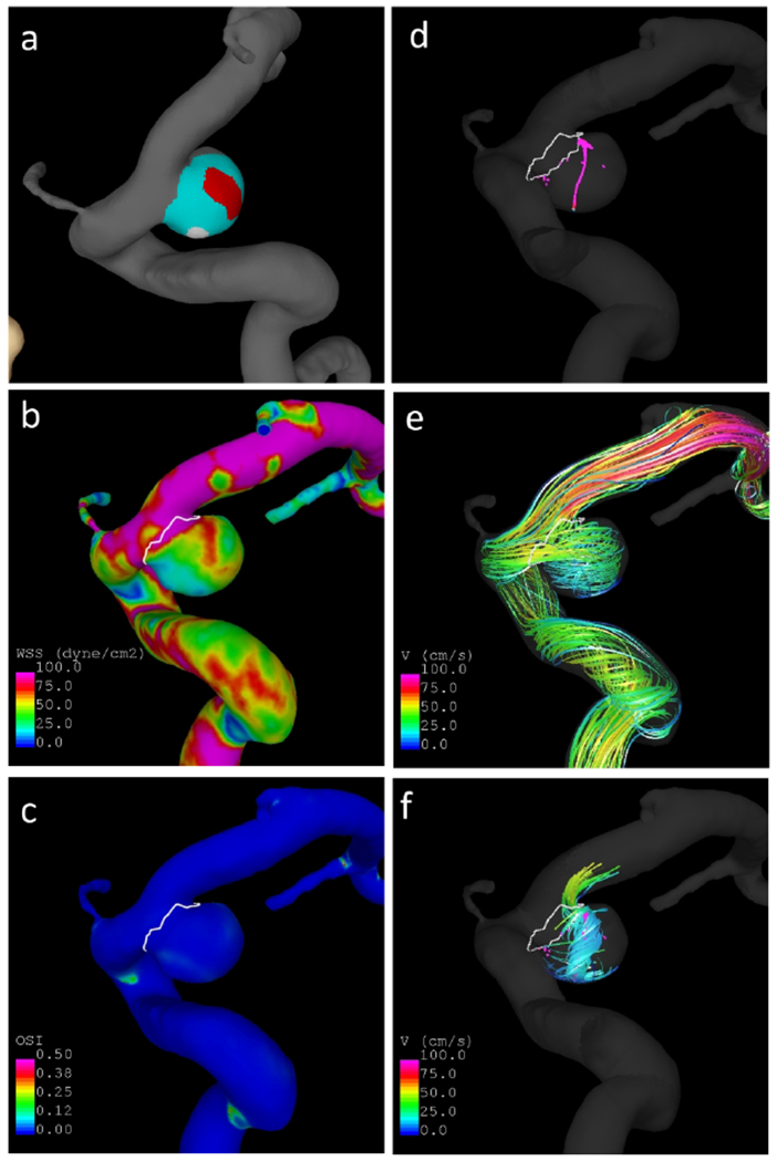
Figure 2.

Example aneurysm with thin and hyperplastic regions: a) wall regions (red=thin, white=hyperplastic, cyan=normal looking), b) wall shear stress, c) oscillatory shear index, d) vortex core-lines, e) flow streamlines, f) swirling around vortex core-lines.
