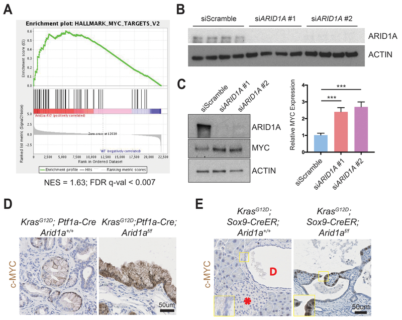
Figure 5.
ARID1A loss induces gene signatures associated with MYC activity.
(A) Gene set enrichment analysis of RNA-seq of Ptf1a-Cre; Arid1af/f (CA) pancreata using the “Hallmark” gene sets demonstrated that the MYC target gene signature was the most upregulated signature in CA mice. NES = normalized enrichment score; FDR = false discovery rate.
(B) Western blot confirmed that siARID1A #1 and #2 were effective in knocking down ARID1A in human pancreatic ductal epithelial (HPDE) cells.
(C) Western blot and quantification demonstrating that ARID1A knockdown induced MYC expression in human pancreatic ductal epithelial (HPDE) cells. n = 8, ***P < 0.001.
(D) PanIN in KrasG12D; Ptf1a-Cre; Arid1a+/+ mice had patchy c-MYC expression, while cysts in KrasG12D; Ptf1a-Cre; Arid1af/f mice stained intensely for c-MYC.
(E) In KrasG12D; Sox9-CreER; Arid1a+/+ mice, acinar, duct (“D” in duct lumen), and islet (asterisk) cells had very little c-MYC staining, while cysts from KrasG12D; Sox9-CreER; Arid1af/f mice expressed some c-MYC.

