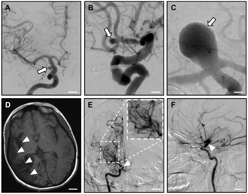
Figure 2: Intracranial Aneurysms (IA) and Moyamoya Disease (MMD).
A-C: Intracranial aneurysms. Computed tomography (CT) angiography studies demonstrate cerebral aneurysms (arrows) of the supraclinoid portion of the internal carotid artery (A); the anterior communicating artery (B); and the basilar artery (C). Scale bars: (A) 0.7 cm; (B) 0.5 cm; (C) 0.2 cm.
D-E: Moyamoya disease. MR imaging (D) shows right-sided encephalomalacia (loss of brain tissue after repeated strokes; arrowheads) above the middle cerebral artery, arising from stenosis/occlusion of the right internal carotid artery. Anterior-posterior (E) and lateral (F) CT angiography reveals occlusion of the right internal carotid artery (arrowhead) with a 'puff-of-smoke' network of collateral vessels (inlay in E). Scale bars: (D) 2 cm; (E, F) 1.3 cm. Images courtesy of Drs. Charles Matouk and Branden Cord (Department of Neurosurgery, Yale School of Medicine and Yale New Haven Hospital).

